When I began writing an article on the physical qualities which are related to being an ultra-fast skater, I began with ankle dorsiflexion…which resulted in a deep dive…and, now, have found that the article would be too long to cover anything else. Without further ado, here’s a write-up on ankle dorsiflexion and on-ice performance.
Ankle Dorsiflexion and Off-ice Injury Risk
Having adequate motion in the ankle is key to efficient, safe movement in sport. While each sporting context differs, high-level athletes must have sufficient range of motion to (1) get into the most advantageous body positions for their sport, (2) produce safe and efficient movement from these positions, and (3) absorb incoming forces from these positions. Before getting into how ice hockey is different from land-based athletic endeavors, let’s first discuss a bit of research. Limited ankle dorsiflexion has been linked to several lower extremity injuries in athletic populations, including ACL tears [1, 2], Achilles ruptures and tendinopathy [3, 4], patellar tendon pain [5-7], and ankle sprains [8]. Poor performance during complex tasks which require substantial ankle dorsiflexion has also been associated with lower extremity injury risk. A few examples of this phenomenon are provided below.
For example, in a large study of 551 NCAA Division II collegiate athletes by Hartley et al. (2018), poor anterior reach performance on the Y-Balance test was associated with likelihood for subsequent ankle sprain injury [9].
Mauntel et al. (2013) observed unfavorable single-leg squat kinematics in individuals with limited ankle dorsiflexion range of motion (ROM). The researchers had a group of individuals perform single-leg squats and categorized them as having medial knee displacement, or not [10]. Those identified to have medial knee displacement during the squat also presented with limited ankle dorsiflexion ROM. The researchers suggest that the limited ankle dorsiflexion ROM resulted in neuromuscular compensation during the single-leg squat pattern, exhibited by the medial knee displacement (i.e. knee-valgus), and coinciding lower levels of gluteal muscle activation relative to hip adduction activation [10]. Significant medial knee displacement (i.e. knee-valgus) is a well-known risk factor for numerous lower extremity injuries [39, 40, 46].
In a cohort of 306 basketball and floorball athletes, Raisan et al. (2017) recently found that athletes with high frontal knee projection angles (i.e. knee valgus) during single-leg squats were 2.7x to sustain an injury in the lower extremity and ankle within the following 12 months, compared with athletes with low frontal knee projection angles [11]. Similarly, having a high frontal knee projection angle was associated with a 2.4x increased risk for sustaining an injury to the ankle [11]. Lower extremity frontal plane projection angles (FPPA) are frequently used as a measure of dynamic knee valgus during functional tasks, such as the single-leg squat [38]. Increased dynamic knee valgus is observed in people with knee pathologies including patellofemoral pain [39] and ACL injury [40].
It’s becoming increasingly clear that, when weight-bearing ankle dorsiflexion ROM is limited, the range of motion of the knee and the trunk in the sagittal plane is reduced, producing compensation forces in the frontal and transverse planes, which could lead to these types of injuries [10-14]. In fact, a recent meta-analysis of 17 studies by Lima et al. (2018) demonstrated an association between ankle dorsiflexion ROM and dynamic knee valgus [15]. The authors concluded that lack of ankle dorsiflexion ROM may be related to harmful movement patterns of the lower limbs [15]. There are various other studies which indicate this notion, including recent works by Malloy et al. (2015), Dowling et al. (2018), and Howe et al. (2019). For the research nerds (like myself), I’ll touch on each of these studies briefly, and then we can move on to on-ice ankle dorsiflexion data in ice hockey athletes.
Malloy et al. (2015) observed that soccer players who presented with reduced ankle dorsiflexion ROM performed a bilateral landing task with greater peak knee abduction angles (i.e. knee valgus) [16], which is a significant risk factor for anterior cruciate ligament (ACL) injury [17].
In a single leg jump landing task, Dowling et al. (2018) found that male athletes with limited ankle dorsiflexion had decreased knee flexion at initial contact, maximal knee flexion, as well as total excursion [18]. Ultimately, Dowling et al. (2018) concluded that restricted ankle dorsiflexion ROM may alter single-leg landing mechanics which may predispose the athlete to injury [18].
Howe et al. (2019) recently found that recreational athletes with limited ankle dorsiflexion ROM tended to land with greater ankle plantarflexion and knee extension at initial contact, alongside reduced ankle dorsiflexion and knee flexion at the maximum flexion point during bilateral drop-landing tasks [19]. Additionally, the recreational athletes with limited ankle dorsiflexion ROM exhibited frontal-plane compensations via increased lower-extremity peak frontal plane projection angle (FPPA), [19]. As touched on previously, high FPPA indicate knee valgus [38]. Knee valgus [42] and stiff landings (also observed in this study) [41] have been associated with non-contact lower extremity injury risk. The authors speculate that the observed movement compensation strategies may allow athletes to better manage the vertical forces experienced during landings, but at the expense of increasing lower extremity injury risk during landing activities [19].
Ankle Dorsiflexion and Skating Speed
So, ankle dorsiflexion ROM appears to matter (a lot) for athletes that perform on land. What about athletes that make their living on-ice? I have yet to come across any peer-reviewed literature that investigates ankle dorsiflexion ROM and injury risk in ice hockey athletes, but there’s a tiny bit of research which looks at ankle dorsiflexion ROM in the skate and on-ice prowess. Let’s go through that.
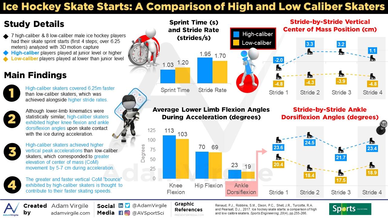
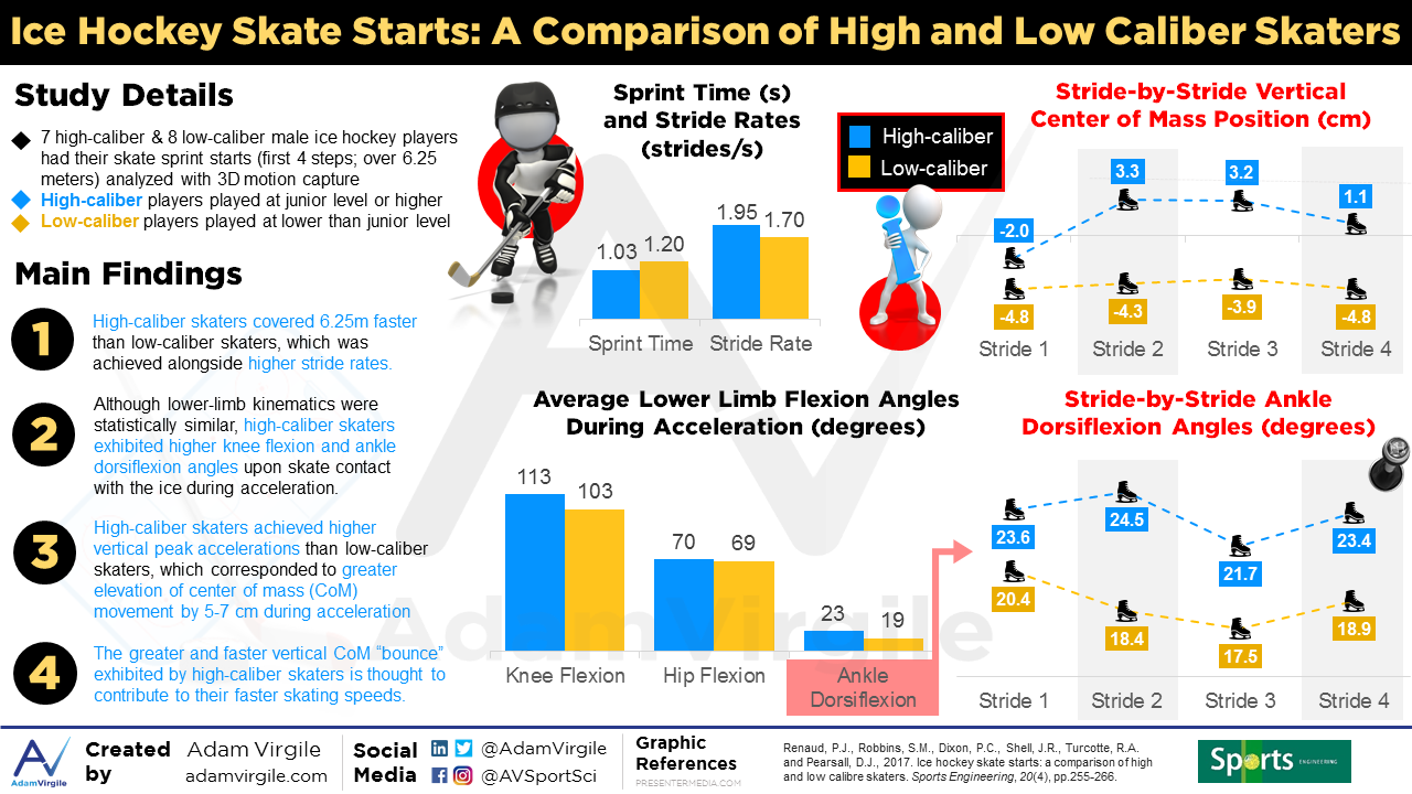
Recently, Renaud et al. (2017) took a look at the strides of 7 high-caliber skaters who played at the junior level or higher, and 8 low-caliber skaters who played below junior level [20]. They used a 10-camera 3-D motion capture system to identify differences in joint kinematics during maximal skating sprint starts on-ice.
High-caliber skaters performed the skating start in a significantly shorter time than low-caliber skaters, despite no differences in lower body power (as estimated from long jump distances). High-caliber skaters generated significantly greater peak accelerations in the vertical direction (9.3-10.6 m/s2 for high-caliber vs. 3.8-8.4 m/s2 for low-caliber skaters), which was related to greater center of mass (CoM) vertical “bounce” by 5-7 centimeters during acceleration [20]. Although both groups displayed significant levels of ankle dorsiflexion (20-30 degrees), dorsiflexion angles were also greater in the high-caliber skaters (the difference wasn’t statistically significant, but there’s a clear difference when viewing the data: see below). This relationship makes sense because higher degrees of ankle dorsiflexion would lead to a greater pre-dorsiex position of the ankle, which, in turn may contribute to a greater ‘‘plantar coil reflex’’ action and yield greater and faster vertical CoM flight. To simplify, high-caliber skaters accelerated faster and had greater vertical “bounce” and ankle dorsiflexion while doing it.
Upjohn et al. (2008) had 5 high-caliber (McGill men’s varsity ice hockey team) and 5 low-caliber (general public) ice hockey players perform 3x 1-minute maximal skating speed intervals on a skating treadmill. Data was captured using four 1.33- megapixel digital video camcorders (Cannon Optra 200 MC), recording at 60 frames per second. Similar to the observations by Renaud et al. (2017), the high-caliber skaters achieved higher skating velocities than their low-caliber counterparts [21]. Large differences in ankle-foot kinematics were observed, with high-caliber skaters showing greater ankle dorsiflexion during weight acceptance and greater rates of plantarflexion during propulsion [21].
Robbins et al. (2018) had 8 high-caliber male ice hockey players (recruited from the university varsity team, who had played at least at the major junior level) and 8 low-caliber male ice hockey players (recruited from local recreational teams and played hockey at levels lower than major junior) perform 5x forward skating sprints over 19.1m [22]. Data was captured via 10-camera system sampled at 240 frames per second (Vicon Motion Systems Ltd., Oxford, UK; eight MX3+ cameras, and two T40S cameras). The data collection began when the athletes entered the recording area, after 6.1m of acceleration [22].
Greater change in ankle dorsiflexion during glide to plantarflexion during push-off (i.e., greater sagittal ankle excursions) were related to faster speeds in the low-caliber skaters, but this relationship did not exist in high-caliber skaters [22].
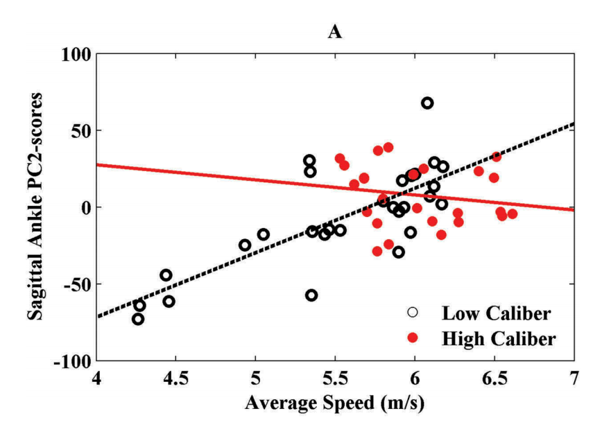
If you take the pool of skaters, it’s clear that greater sagittal ankle excursions (i.e. PC2-scores) segregate the faster skaters from the slower skaters, as well as the high-caliber skaters from the low-caliber skaters [22]. In their conclusion, Robbins et al. (2018) state that “increased excursion from ankle dorsiflexion during glide to plantarflexion during push-off” should be recommended to players and coaches because it might lead to improved skating performance [22].
As we will soon see in the following skate design literature, increasing the capacity to dorsiflex the ankle does not necessarily result in increased skating speed, even when initial ankle dorsiflexion capacity is limited.
Skate Design Impact on Ankle Motion
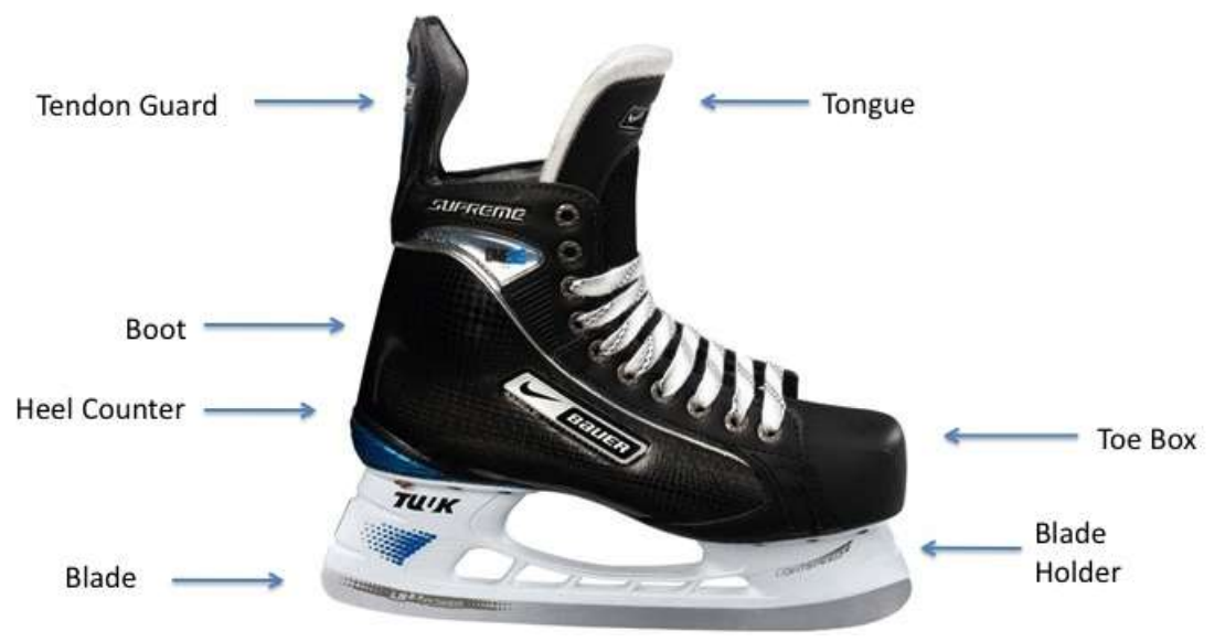
Skate fit is a huge deal in ice hockey performance. If the skate is too loose, there will be foot slipping within the boot, which will result in instability and reduce push-off power [23]. If the skate is too tight, mobility will be limited, also reducing push-off power. An optimal “snug” fit allows for a better foot/skate boot interface, which in turn increases the perception of foot orientation and kinaesthetic feedback. This is because a good fit likely helps to provide afferent information to the central nervous system (CNS), which optimizes kinematic adjustments during skating performance, while also allowing for the necessary mobility to occur to optimize power output [23, 24].
In recent history, skate manufacturers have toyed around with modifications of traditional skate design in order to put players at a more favorable biomechanical advantage via increasing ankle range of motion. This revolutionary concept of increasing ankle range of motion arose with the invention of the klapskate in the sport of speed skating [25, 26]. The klapskate is designed with a hinge under the anterior part of the skate boot [27, 43]. The studies on the effects of the klapskate on speed skating performance have revealed the importance of increased ankle range of motion that the klapskate provides for improvements in speed skating performance [28].
Various kinematic analyses using ice hockey skate models have suggested that the type of hockey skate an athlete wears can affect the range of motion of the ankle and subtalar joint during the skating stride [27, 29-32]. Dewan et al. (2004) were quick to point out how crucial proper skate fit and comfort can be to foot-ankle kinematics inside the boot [33]. The impact of allowing for dynamic changes of the foot and ankle structures to occur within the boot was clear, and skate manufacturers took note. Skate manufacturers began modifying traditional skate design by removing the rear tendon guard (i.e. Achilles guard) or making this portion of the skate more flexible. Research on these modifications will be discussed below.
IMAGE OF SKATE FROM STIDWILL 2009 [34].
Hellyer et al. (2016) evaluated how 8 highly skilled male hockey players with Canadian Interuniversity Sport or professional hockey experience skated on a skating treadmill at varying velocities using two different skate designs [32]. One design was the traditional ice hockey skate, and the other was the Easton Mako skate which had a flexible rear tendon guard [32]. Ankle dorsiflexion range of motion was lower, but plantarflexion range of motion was higher in the Easton Mako skate with a more flexible rear tendon guard (“modified”), compared with the traditional skate [32].
This resulted in higher plantarflexion angular velocities during skating with the modified Easton Mako skate and, ironically, knee extension, hip extension, and hip abduction angular velocities followed suit. Increased range of motion at each of these joints, as well as increased angular velocities at each of these joints, were related with increased stride width, length, rate, and velocity [32]. In other words, and I quote the authors, “this difference together with the increased range of motion exhibited by the use of this new skate design indicates that not only was the range of motion larger while wearing the Easton Mako, but also the movement was occurring at a faster rate. Assuming the recovery phase of the stride does not change while using this new skate design, the increase in angular velocity will increase the stride rate, which is a characteristic of faster skaters,” [32].
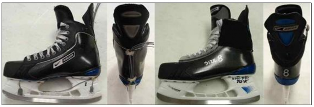
The next few studies I’m going to discuss come out of McGill University [27, 31, 35, 36]. These studies compare kinetics and kinematics in athletes wearing a pair of regular skates with the kinetics and kinematics wearing a modified pair of skates. The skate designs used in the following studies [27, 31, 35, 36] are described below:
Regular skate: A standard Bauer One95 ice hockey skate
Modified skate: A One95 ice hockey skate including a modified flexible Achilles tendon guard and a modified eyelet placement at the metatarsal guard allowing for increased plantarflexion and dorsiflexion
One of the McGill studies was conducted by Le Ngoc (2013). During acceleration, values of impulse, peak and average vertical force, work, and power were all lower when wearing the modified skate design during sprinting, compared with the regular skate [35]. There were no statistically significant differences in stride rates or contact times during acceleration.
Generally, a double peak pattern is observed in the vertical force applied to the ice during the contact phase [35]. These two peaks represent (1) the weight acceptance upon skate touchdown to the ice and (2) the propulsion of the skate from the ice, respectively. In the study by Le Ngoc (2013), the peak vertical force consistently occurred at the propulsion phase of the stride in the modified skate, whereas the peak vertical force consistently occurred at the weight acceptance phase of the stride in the regular skate [35]. More specifically, the peak vertical force occurred at 80-81% of stride completion in the modified skate and at 45-60% of stride completion in the regular skate. Impulse, work, and power values are all calculated from the vertical force being applied to the ice during weight acceptance and propulsion. According to Le Ngoc (2013), “the modified skate seemed to allow the ankle to be in a more dorsiflexed position which might have resulted in a smoother weight acceptance by having the blade at a more horizontal level as it contacts the ice. This might result in less forceful impact which results in less vertical force registered by the strain gauge system during weight acceptance. That may, in turn, explain the lower values of impulse, work and power as well as the consistent peak vertical force during the propulsion phase of the stride,” [35].
In a similar study, Culhane (2012) also observed lower values of impulse, work and power when wearing the modified skate, compared with the regular skate [36]. These observations are in line with those aforementioned in a similar study by Le Ngoc (2013). Like Le Ngoc (2013), the reduced values of impulse, work, and power observed by Culhane (2012) could have been due to less forceful impact upon ice contact via the increased allowable ankle dorsiflexion provided by the modified skate design [36]. Also similar to Le Ngoc (2013), Culhane (2012) did not observe statistically significant differences in contact times or stride rates during acceleration between skate models [36].
Forget (2013) performed a similar investigation in 14 healthy young males (19-29 years of age) varying in hockey skill from high to low caliber [27]. Although the modified skate design used was identical to that of Le Ngoc (2013) and Culhane (2012), the on-ice protocol was very different in the study by Forget (2013). In contrast to the linear sprint protocols of Le Ngoc (2013) and Culhane (2012), Forget (2013) had the skaters perform explosive stop-and-go task on-ice [27]. It’s important to note that the natural kinetics and kinematics involved with the stop-and-go task (Forget, 2013) are very different from “go” from a dead stop (Le Ngoc, 2013; Culhane, 2012), and thus, the results from Forget (2013) should be interpreted with this difference in mind.
As one might expect, the high-caliber skaters were significantly faster their low-caliber counterparts but there were no differences between groups in the vertical force or impulse variables [27].
Just as we saw from Le Ngoc (2013) and Culhane (2012), there were no differences in on-ice performance (contact times, stride rates, time to completion) between skate models. Greater vertical force and impulse values were observed when using the regular skate, compared with the modified skate [27]. Once again, this data might speak toward the advantageous lessened impact upon weight acceptance via increased available ankle dorsiflexion range of motion when using the modified skate, compared with the regular skate. This extrapolation on the data is purely conjecture, but also makes physiological sense. Average force values were higher in the modified skate in the heel and mid foot region, but lower in the toe region, during on-ice sprinting [27]. While it’s difficult to determine precisely why this would be, these results suggest that one should appreciate that different skate models will alter force application strategies on the ice [27].
In another study by Robert-Lachaine et al. (2012),10 healthy adult male hockey players performed maximal forward skating sprints and crossovers in each direction, on-ice [31]. The skaters performed these sprints on two occasions: once wearing a pair of regular hockey skates, and once wearing a pair of modified skates which were manufactured with the same characteristics as the regular hockey skates, but with lighter and more flexible tongues [31]. Other alterations to the modified pair of skates were (1) the eyelets were slightly raised away from the skate, and (2) the tendon guard was cut down to the calcaneus level and attached with an elastic band [31].
While there wasn’t a statistically significant difference in ankle dorsiflexion range of motion between skate types, increased plantarflexion (3-5 degrees) and total dorsiflexion-plantarflexion (5-10 degrees) range of motion was observed using the modified skate, compared with the regular skate [31]. Despite (1) a slightly increased stride rate, (2) increased impulse, (3) increased power, and (4) increased work during the skating tasks using the modified skate, there were no differences in task completion times.
This is interesting, and appears to conflict with the findings of the studies previously noted. However, the authors of this study report that there was particularly high variability regarding the skating kinetics in the group using the modified skate; half of the skaters had more favorable skating kinetics using the modified skate, while the other half of the skaters displayed equal or inferior skating kinetics, compared with the regular skate [31].
Now, why is this relevant, you ask?
Pragmatically, these results indicate that several skaters may not have been able to take full advantage of the skate design modifications in such a short period of time. While some skaters can adapt quickly to novel skating mechanics utilizing more favorable biomechanical positions, others need more time to learn how to take advantage of this positioning. It’s possible that a few skaters exhibited dramatically higher work, power, and impulse values using the modified skate upon weight acceptance due to lack of familiarization (i.e. weren’t able to increase “smoothness” of skate touchdown via increased available ankle range of motion), whereas the other half of skaters produced lower kinetic values because they were able to adjust more quickly to the modified skate design [31].
Skate Lacing Technique and Ankle Motion
USA Hockey provides a nice tutorial on how to get dressed for ice hockey for parents and young skaters (clip below); 2:00-2:30 addresses skate lacing. Parents are taught to tie their kids’ skates through the top eyelet because the ankle inside the skate will feel tight and secure [44, 45]. As discussed, a major drawback of this strategy is the reduction of accessible ankle motion. Consequently, kids become accustomed to limited ankle range of motion within the boot from a very young age.
A common strategy to increase ankle motion without altering the skate is to leave the top eyelet undone, or without lace. While the increased lateral (side-to-side) security is important to consider, the increased sagittal (front-and-back) mobility may allow for the athlete to (1) skate faster during propulsion, and (2) have greater control upon weight acceptance.
Caution: Extrapolation on bad science, ahead! You’ve been warned.
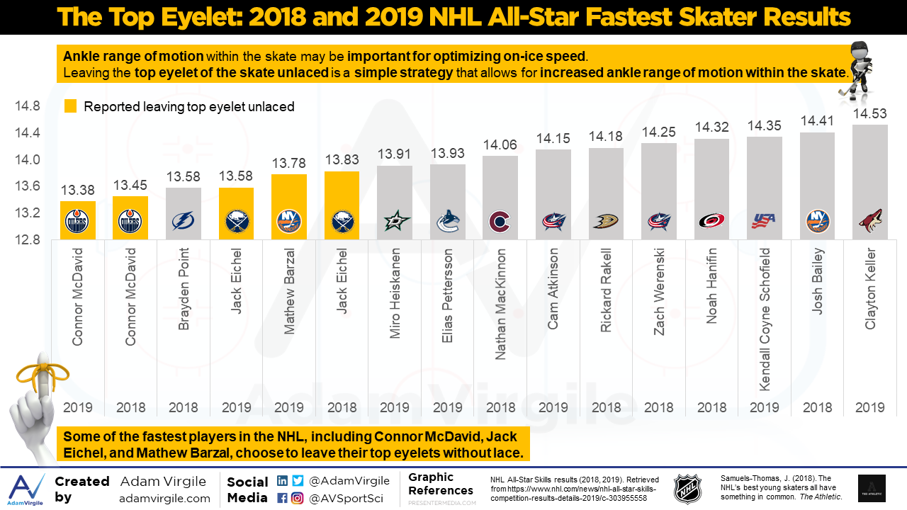
The trend of leaving the top eyelet undone has emerged among younger elite ice hockey athletes because, early on in their careers, they “found it cool to leave the top lace unlocked in order to have the tongues flop down on the skates,” according to Dr. Lockwood in her interview with The Athletic [37]. Some of these iconic young players include Connor McDavid, Jack Eichel, Johnny Gaudreau, Auston Matthews, Brady Tkachuk, Shayne Gostisbehere, Mathew Barzal, Taylor Hall (second eyelet untied instead of top), Jason Zucker, Mark Scheifele, Anthony Duclair, Morgan Rielly [37]. Although anecdotal, some of these players are fast skaters. And when I say fast, I mean REALLY fast. Most of my opinions are structured around loads of peer-reviewed evidence, but I don’t have any peer-reviewed data that speaks to this claim. However, I do know that Connor McDavid won the Fastest Skater competition at the All-Star game festivities in 2017… and 2018….and for the third consecutive year in 2019. Jack Eichel came in second in 2019, and Johnny Gaudreau was too busy winning the Puck Control competition.
At the time these players began unlacing the top eyelet they were, likely, unaware of the huge biomechanical advantage that could be unlocked, according to the research noted above. However, the aforementioned players have spent years, if not decades, skating in this way, which gave them time to adapt their skating styles and develop effective skating technique using the additional ankle range of motion that this strategy provides.
As we saw in the aforementioned research, increasing ankle range of motion through modified skate design did not lead to faster skating speeds, but the athletes who participated in the studies only used the modified skates once, or a mere handful of times. They were not given enough time to familiarize themselves with the skate and figure out how to effectively utilize all of the potential biomechanical design benefits. Again, given the inferences from previous research, I believe that athletes that alter skate lacing strategy, such as leaving the top eyelet unlaced, must also learn how to safely and effectively utilize the gained ankle range of motion in order to maximize the biomechanical benefits that such a strategy provides. It’s important to note that on-ice maneuvers are multifaceted and complex. Not only must a player accelerate, but he or she must also decelerate, stop, change direction, or turn in response to game cues. While this article focuses on on-ice acceleration and sprinting performance from a dead stop, consideration for how increased ankle motion within the boot could affect the safety and efficiency of other skating maneuvers is of paramount importance.
Summary
- Limited ankle dorsiflexion has been associated with a range of lower extremity injuries in land-based sports, but there’s a paucity of research in this area in ice hockey athletes.
- High-caliber skaters are faster than their lower-caliber counterparts.
- High-caliber skaters typically utilize greater ankle dorsiflexion-plantarflexion ranges of motion during acceleration and sprint skating than their lower-caliber counterparts.
- Research comparing traditional or “regular” skate design to skates that have been modified to allow for increased ankle range of motion (via inclusion of a modified flexible Achilles tendon guard and a modified eyelet placement at the metatarsal guard) suggests that ankle dorsiflexion and plantarflexion range of motion is increased in modified skates, but that skating speed does not follow suit.
- In modified skates (skates that include a modified flexible Achilles tendon guard and a modified eyelet placement at the metatarsal guard), kinetic variables, such as power, impulse, and work are typically reduced, suggesting (potentially) a more controlled foot-ankle complex upon weight acceptance, which could reduce lower-extremity injury risk.
- Leaving the top eyelet of the skates unlaced is becoming a more common occurrence, albeit probably not for any biomechanical advantage rationale.
- Leaving the top eyelet of the skates unlaced may help to further promote ankle dorsiflexion and plantarflexion range of motion, without any necessary skate modifications.
For a review of the off-ice physical qualities that are associated with on-ice skating speed and on-ice success, you can go here.
Reference
- Wahlstedt, C. and Rasmussen-Barr, E., 2015. Anterior cruciate ligament injury and ankle dorsiflexion. Knee Surgery, Sports Traumatology, Arthroscopy, 23(11), pp.3202-3207.
- Amraee, D., Alizadeh, M.H., Minoonejhad, H., Razi, M. and Amraee, G.H., 2017. Predictor factors for lower extremity malalignment and non-contact anterior cruciate ligament injuries in male athletes. Knee Surgery, Sports Traumatology, Arthroscopy, 25(5), pp.1625-1631.
- Pope R, Herbert R, Kirwan J. Effects of ankle dorsiflexion range and pre-exercise calf muscle stretching on injury risk in Army recruits. Australian Journal of Physiotherapy. 1998;44(3):165-72. PubMed PMID: 11676730. Epub 2001/10/26. Eng.
- Rabin, A., Kozol, Z. and Finestone, A.S., 2014. Limited ankle dorsiflexion increases the risk for mid-portion Achilles tendinopathy in infantry recruits: a prospective cohort study. Journal of foot and ankle research, 7(1), p.48.
- Malliaras P, Cook JL, Kent P. Reduced ankle dorsiflexion range may increase the risk of patellar tendon injury among volleyball players. Journal of Science and Medicine in Sport. 2006 Aug;9(4):304-9. PubMed PMID: 16672192. Epub 2006/05/05. Eng.
- Witvrouw E, Lysens R, Bellemans J, Cambier D, Vanderstraeten G. Intrinsic risk factors for the development of anterior knee pain in an athletic population. A two-year prospective study. American Journal of Sports Medicine. 2000 JulAug;28(4):480-9. PubMed PMID: 10921638. Epub 2000/08/02. Eng.
- Backman, L.J. and Danielson, P., 2011. Low range of ankle dorsiflexion predisposes for patellar tendinopathy in junior elite basketball players: a 1-year prospective study. The American journal of sports medicine, 39(12), pp.2626-2633.
- Drewes LK, McKeon PO, Kerrigan DC, Hertel J. Dorsiflexion deficit during jogging with chronic ankle instability. Journal of science and medicine in sport / Sports Medicine Australia. 2009 Nov;12(6):685-7. PubMed PMID: 18835218. Epub 2008/10/07. Eng.
- Hartley, E.M., Hoch, M.C. and Boling, M.C., 2018. Y-balance test performance and BMI are associated with ankle sprain injury in collegiate male athletes. Journal of science and medicine in sport, 21(7), pp.676-680.
- Mauntel, T.C., Begalle, R.L., Cram, T.R., Frank, B.S., Hirth, C.J., Blackburn, T. and Padua, D.A., 2013. The effects of lower extremity muscle activation and passive range of motion on single leg squat performance. The Journal of Strength & Conditioning Research, 27(7), pp.1813-1823.
- Räisänen, A.M., Pasanen, K., Krosshaug, T., Vasankari, T., Kannus, P., Heinonen, A., Kujala, U.M., Avela, J., Perttunen, J. and Parkkari, J., 2018. Association between frontal plane knee control and lower extremity injuries: a prospective study on young team sport athletes. BMJ open sport & exercise medicine, 4(1), p.e000311.
- Bell-Jenje, T., Olivier, B., Wood, W., Rogers, S., Green, A. and McKinon, W., 2016. The association between loss of ankle dorsiflexion range of movement, and hip adduction and internal rotation during a step down test. Manual therapy, 21, pp.256-261.
- Dill, K.E., Begalle, R.L., Frank, B.S., Zinder, S.M. and Padua, D.A., 2014. Altered knee and ankle kinematics during squatting in those with limited weight-bearing–lunge ankle-dorsiflexion range of motion. Journal of athletic training, 49(6), pp.723-732.
- Ota, S., Ueda, M., Aimoto, K., Suzuki, Y. and Sigward, S.M., 2014. Acute influence of restricted ankle dorsiflexion angle on knee joint mechanics during gait. The Knee, 21(3), pp.669-675.
- Lima, Y.L., Ferreira, V.M.L.M., de Paula Lima, P.O., Bezerra, M.A., de Oliveira, R.R. and Almeida, G.P.L., 2018. The association of ankle dorsiflexion and dynamic knee valgus: A systematic review and meta-analysis. Physical Therapy in Sport, 29, pp.61-69.
- Malloy, P., Morgan, A., Meinerz, C., Geiser, C. and Kipp, K., 2015. The association of dorsiflexion flexibility on knee kinematics and kinetics during a drop vertical jump in healthy female athletes. Knee Surgery, Sports Traumatology, Arthroscopy, 23(12), pp.3550-3555.
- Hewett, T.E., Myer, G.D., Ford, K.R., Heidt Jr, R.S., Colosimo, A.J., McLean, S.G., Van den Bogert, A.J., Paterno, M.V. and Succop, P., 2005. Biomechanical measures of neuromuscular control and valgus loading of the knee predict anterior cruciate ligament injury risk in female athletes: a prospective study. The American journal of sports medicine, 33(4), pp.492-501.
- Dowling, B., Mcpherson, A.L. and Paci, J.M., 2018. Weightbearing ankle dorsiflexion range of motion and sagittal plane kinematics during single leg drop jump landing in healthy male athletes. The Journal of sports medicine and physical fitness, 58(6), pp.867-874.
- Howe, L.P., Bampouras, T.M., North, J. and Waldron, M., 2019. Ankle dorsiflexion range of motion is associated with kinematic but not kinetic variables related to bilateral drop-landing performance at various drop heights. Human movement science, 64, pp.320-328.
- Renaud, P.J., Robbins, S.M., Dixon, P.C., Shell, J.R., Turcotte, R.A. and Pearsall, D.J., 2017. Ice hockey skate starts: a comparison of high and low calibre skaters. Sports Engineering, 20(4), pp.255-266.
- Upjohn, T., Turcotte, R., Pearsall, D.J. and Loh, J., 2008. Three-dimensional kinematics of the lower limbs during forward ice hockey skating. Sports biomechanics, 7(2), pp.206-221.
- Robbins, S.M., Renaud, P.J. and Pearsall, D.J., 2018. Principal component analysis identifies differences in ice hockey skating stride between high-and low-calibre players. Sports Biomechanics, pp.1-19.
- Turcotte, R.A., Renaud, P. and Pearsall, D.J., 2016. Ice Hockey Skate, Stick Design and Performance Measures. In The Engineering Approach to Winter Sports (pp. 311-326). Springer, New York, NY.
- Kekoni, J., Hämäläinen, H., Rautio, J. and Tukeva, T., 1989. Mechanical sensibility of the sole of the foot determined with vibratory stimuli of varying frequency. Experimental brain research, 78(2), pp.419-424.
- Hoshizaki, T. B., Hall, K., & Bourque, R. (1989). Montreal, Canada Patent No.: US. Patent No. 4, 885.
- Madore, C. (2003). Montreal, Canada Patent No.: U. Patent.
- Forget, S., 2014. Comparisons of player calibers and skate models during an ice hockey explosive transitional maneuver (Doctoral dissertation, McGill University Libraries).
- de Koning, J.J., Houdijk, J.H.P., de Groot, G. and Bobbert, M.F., 2000. From biomechanical theory to application in top sports: the klapskate story. Journal of Biomechanics, 33, pp.1225-1229.
- Baig, Z.A., 2011. Functional Mechanical Assessment of Foot and Ankle: Stiffness and Work Production in Ice Hockey Skate Boots (Doctoral dissertation, McGill University Library).
- Fortier, A., 2010. Skating mechanics of change of direction maneuvers in hockey players (Doctoral dissertation, McGill University Library).
- Robert-Lachaine, X., Turcotte, R.A., Dixon, P.C. and Pearsall, D.J., 2012. Impact of hockey skate design on ankle motion and force production. Sports engineering, 15(4), pp.197-206.
- Hellyer, M.R., Alexander, M.J., Glazebrook, C.M. and Leiter, J., 2016. Differences in Lower Body Kinematics during Forward Treadmill Skating Between Two Different Hockey Skate Designs. International Journal of Kinesiology and Sports Science, 4(1), pp.1-16.
- Dewan, C., Pearsall, D. and Turcotte, R., 2008, April. Dynamic pressure measurement about the foot and ankle. In ISBS-Conference Proceedings Archive (Vol. 1, No. 1).
- Stidwill, T.J.L., 2009. Comparison of forward hockey skating kinetics and kinematics on ice and on a synthetic surface by means of a customized force measurement system and electrogoniometry (Doctoral dissertation, McGill University).
- Le Ngoc, C., 2013. Force and Centre of Pressure Measurements During Ice Hockey Skating with a Regular and a Modfied Ice Hockey Skate (Doctoral dissertation, McGill University Libraries).
- Culhane, L., 2012. Acceleration Characteristics of Forward Skating (Doctoral dissertation, McGill University).
- Samuels-Thomas, J. (2018, December 07). The NHL’s best young skaters all have something in common –… Retrieved from https://theathletic.com/696003/2018/12/07/the-nhls-best-young-skaters-all-have-something-in-common-how-they-tie-their-skates/
- Wyndow, N., De Jong, A., Rial, K., Tucker, K., Collins, N., Vicenzino, B., Russell, T. and Crossley, K., 2016. The relationship of foot and ankle mobility to the frontal plane projection angle in asymptomatic adults. Journal of Foot and Ankle Research, 9(1), p.3.
- Willson, J.D. and Davis, I.S., 2008. Utility of the frontal plane projection angle in females with patellofemoral pain. Journal of Orthopaedic & Sports Physical Therapy, 38(10), pp.606-615.
- Myer, G.D., Ford, K.R., Di Stasi, S.L., Foss, K.D.B., Micheli, L.J. and Hewett, T.E., 2015. High knee abduction moments are common risk factors for patellofemoral pain (PFP) and anterior cruciate ligament (ACL) injury in girls: is PFP itself a predictor for subsequent ACL injury?. Br J Sports Med, 49(2), pp.118-122.
- Leppänen, M., Pasanen, K., Kujala, U.M., Vasankari, T., Kannus, P., Äyrämö, S., Krosshaug, T., Bahr, R., Avela, J., Perttunen, J. and Parkkari, J., 2017. Stiff landings are associated with increased ACL injury risk in young female basketball and floorball players. The American Journal of Sports Medicine, 45(2), pp.386-393.
- Koga, H., Muneta, T., Bahr, R., Engebretsen, L. and Krosshaug, T., 2017. ACL injury mechanisms: lessons learned from video analysis. In Rotatory Knee Instability (pp. 27-36). Springer, Cham.
- van Ingen Schenau, G.J., de Koning, J.J. and Gemser, H., 1999. The muscles of speed skating. Handbook of Competitive Speek Skating, pp.78-99.
- City of Boise Parks and Recreation (2016). Idaho IceWorld Ice Pilots Hockey Parent Information Guide [PDF file]. Retrieved from http://www.teamsideline.com/Assets/451/IceWorld/2016-hockey-parent-guide-edit-4.pdf
- Today’s Parent | Feb 5, 2. (2017, February 08). How to help kids tie skates. Retrieved from https://www.todaysparent.com/family/activities/tie-skates/
- Padua, D.A., Bell, D.R. and Clark, M.A., 2012. Neuromuscular characteristics of individuals displaying excessive medial knee displacement. Journal of Athletic Training, 47(5), pp.525-536.
Want to be the first to know when new content is created? Please enter your email address below.